Saturday, September 15, 2012

Hepatomegaly

It is also necessary in addition to locate the lower border of the Liver and the span of the Liver
it is necessary to assess the consistency of the liver ( soft, elatsic, firm, compact, hard), the liver surface ( smooth, protuberant), the tendernss on pressure.


Friday, September 14, 2012

Clubbing

Clubbing causes

Appearance of Clubbing


Bronchieactasis

 
Conditions associated with Bronchiectasis
 
COPD compared with Bronchiectasis
 
 
Symptoms of acute exacerbation of Bronchiectasis
 
Diagnostic testing for Bronchiectasis


Lymphadenopathy

Causes of Lymphadenopathy

Thursday, September 13, 2012

Atrial Fibrillation

 
Atrial Fibrillation Classification


Strategies for Treatment


Medications for Rate and Rhythm control
Selecting Antiarrhythmic
Anticoagulation
 
CHAD2 score


Transverse Myelitis

Criteria for Idiopathic Transverse Myelitis
 
 
Approach to patient with Transverse Myelitis


Cauda Equina


TIA


Pulmonary Hypertension

 
Classifications
Investigations
Right Heart cath for PPH


Pancytopenia


Mechanisms

A useful test to differentiate between the two is the reticulocyte index and the peripheral blood smear.
 
1- Causes
with Hepatosplenomegaly and Atypical Lymphyoctosis


2-Causes without Splenomegaly
-Aplastic Anemia
-Myelodyplasia

Wednesday, September 12, 2012

Mitral Regurgitation


CHF


Outpatient Management
A-First visit

1- Clinical assessment
        a- Current symptoms
        b- Weight
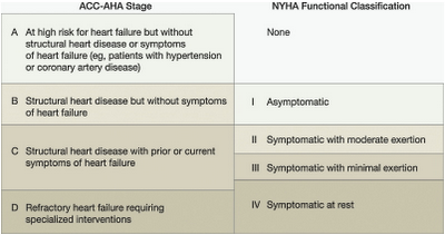
        c- Stage or Functional Class


2- Criteria for Diagnosis 


3- Essential tests and investigation
-ECG
-Basic Labs
-BNP
-Echo for systolic vs. diastolic dysfunction

4-underlying etiology ischemic vs. non ischemic


5-cause of recent decompensation


6-Treatment


B-subsequent visits
f/u clinical    
symptom  signs


Medical Records points


Criteria for Clinical stability of Heart Failure


Thursday, August 23, 2012

Liver disease

The record
The patient is icteric, pigmented. He has clubbing, leuconychia, palmar erythema and dupuytren's contracture and several spider naevi.There is or there is no flapping tremor of the hands. There is scratch marks on the forearms and back, and there is purpura.  There is gynaecomastia, scanty hair and his testes are small.
also he has generalized swelling of the abdomen and the umblicus is everted there is distended abdominal wall veins in which the flow is away from, the umbilicus.The liver is palpable at  .. cm below the right costal margin with a span of about     cm indicating hepatmegaly and 3 cm splenomegaly.  The flanks are dull to percussion but the center is resonant. The dullness is shifting and fluid thrill is present or not  present so he has ascites.  ankle edema is also present .
The diagnosis is likely to be cirrhosis of the liver with portal hypertension.

Sunday, August 5, 2012

Systolic Murmurs

1- Pansystolic murmur : extends throughout systole, begining with the first heart sounds, going right up to the second heart sound.
Occur when a ventricle leaks into a lower pressure chamber or vessel.
Causes include
Mitral Regurgitation , Tricuspid regurgitation and VSD.


Record Description of Mitral Regurgitation
The pulse is regular at 80 beats per min. The venous pressure is not raised and there is no Ankle edema. The apex beat is thrusting ( volume overload) in the sixth inetrcostal space in the anterior axillary line, and there is or there is no thrill.
There is or there is no left parasternal heave. The first heart sound is soft, and there is a third heart sound. There is loud pansystolic murmur at the apex, radiating to the axilla.
The Diagnsois is Mitral Regurgitation with signs of of pulmonary hypertension.


2- Midsystolic Murmur 
Like Aortic stenosis, pulmonary stenosis or hypertrophic cardiomyopathy or pulmonary flow murmur of ASD.
Caused by Trubulent flow through the aortic or pulmonary valve orifices or by greatly increased flow through a normal sized orifice or outflow tract.


Record Desciprtion of Aortic stensois
The pulse is regular at 70 beats per minutes, of small volume and slow rising. The venous pressure is not raised. The apex beat is palpable  1 cm to the left of the midclavicular line in the fifth intercostal as a forceful sustained heave. There is a systolic thrill or there is not thrill palpable over the aortic area radiating into the neck, and the aortic secod sound is soft.
The diagnosis is aortic stenosis.